
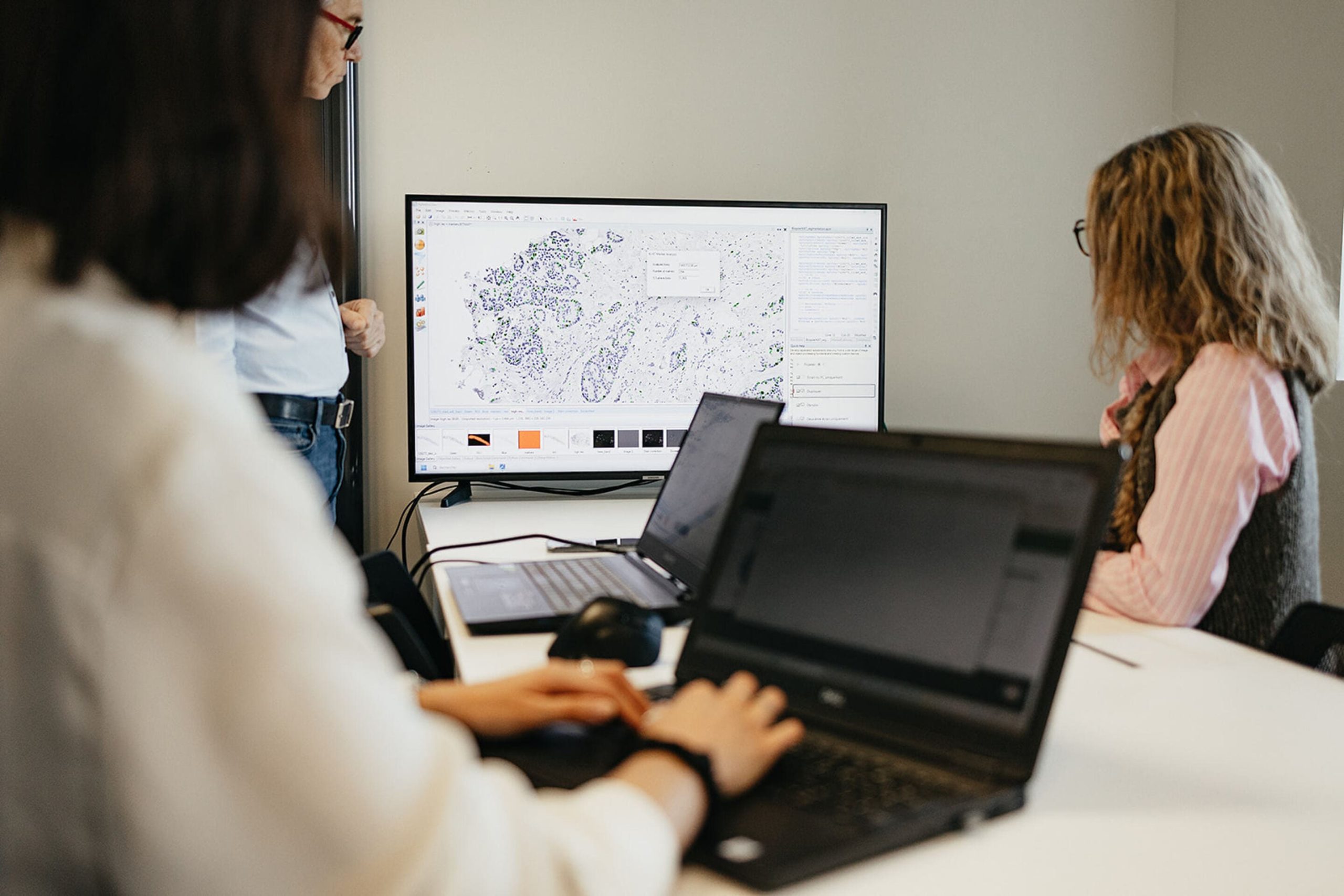
Stereology Analyzer
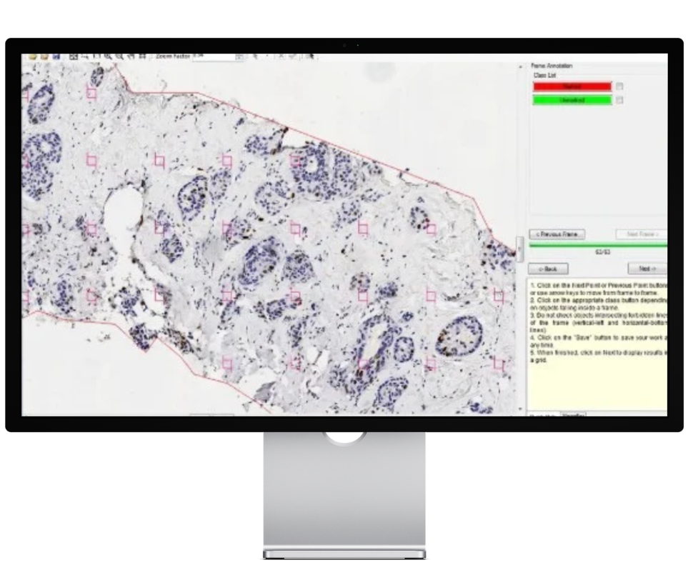
Stereology Analyzer
Turn Images into Quantitative Data
Stereology Analyzer helps transform microscopy images into meaningful scientific measurements.
With Stereology Analyzer, you can:
- Export results for statistical analysis
- Estimate 3D characteristics from 2D images
- Obtain accurate and unbiased measurements
- Analyze large digital slides and microscopy datasets
- Reduce manual counting efforts
- Improve result reproducibility
- Validate automated image analysis and AI algorithms
Applications
- Cell counting and characterization
- Tumor quantification
- Vascular density studies
- Tissue and histopathology analysis
- Grain size analysis
- Inclusion quantification
- Porosity measurements
- Boundary characterization
- Pore analysis
- Phase distribution studies
- Rock structure characterization
Key Features
- Flexible grids: Points, lines, frames, squares, and circles
- Custom ROIs: Rectangles, ellipses, freehand, and custom shapes
- Large image support: TIFF, Tiled TIFF, and JPEG
- Automatic measurements: Volume fraction, numerical density, area estimations, and other stereological metrics
- Data export: Excel®, Word®, and statistical software
Quantify 3D Structures Reliably from 2D Images
Researchers often need to quantify 3D structures such as cells, vessels, tumors, pores, or grains from 2D microscopy and histology images. Achieving accurate and reproducible measurements can be difficult with conventional image analysis methods.
Stereology Analyzer enables accurate and reproducible quantification of 3D structures from 2D images using established stereological methods.


Your Challenges
Obtaining Reliable Quantitative Measurements
Many image analysis workflows struggle to accurately quantify complex structures such as tumors, vessels, cells, pores, or grains, especially when image quality or biological variability limits automation.
Working with Large Histological or Microscopy Images
Whole-slide images and high-resolution composite images often contain large amounts of information that are difficult to analyze consistently and efficiently.
Ensuring Statistical Validity
Manual observations can introduce bias and variability, making it difficult to generate reliable quantitative data for research, regulatory submissions, or publications.
Validating Automated Analysis Methods
Many research groups require a trusted reference method to validate machine learning or image analysis algorithms before clinical or industrial deployment.
The ADCIS Solution
Stereology Analyzer integrates proven stereological and statistical methods into an intuitive platform for digital image analysis. Using interactive grids and ROIs, it enables reliable and reproducible estimation of structural parameters from 2D images.
Scientifically Validated Stereological Methods
The software applies long-established stereological techniques recognized by the scientific community for estimating the characteristics of 3D structures from 2D sections and projections.
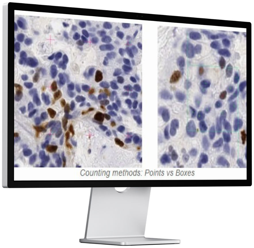
Flexible Grid-Based Analysis
Users can select from multiple grid types, including :
Points – Lines – Frames – Squares – Circles & Advanced grid configurations for specialized measurements
Allowing adaptation to specific research protocols and quantification objectives.
Region-of-Interest (ROI) Management
Researchers can define one or multiple ROIs and focus analysis only on relevant structures,
improving measurement precision and workflow efficiency.
Automated Statistical Calculations
Once structures of interest have been identified, the software automatically performs the stereological calculations
required to estimate quantitative parameters and generate reproducible results.
Use-cases Applications
- Cell counting and density estimation
- Tumor quantification
- Blood vessel analysis
- Placental and tissue structure analysis
- Histopathology research
- Grain size measurement
- Porosity analysis
- Inclusion characterization
- Boundary quantification
- Pore characterization
- Mineral phase quantification
- Rock microstructure analysis
Key Benefits
- Reliable quantitative analysis from 2D images.
- Trusted stereological methods.
- Support for large microscopy images.
- Reduced bias and improved reproducibility.
- Validation of image analysis and AI tools.
- Flexible for diverse research needs.
Who Is It For?
Stereology Analyzer is designed for:
- Research laboratories
- Academic institutions
- Pathology departments
- Pharmaceutical companies
- Medical research organizations
- Materials science laboratories
- Geological research centers
That require robust, statistically sound quantification of microscopic structures.
In One Sentence
Stereology Analyzer enables researchers to obtain accurate and reproducible 3D measurements from 2D microscopy
and histology images using scientifically validated stereological methods.