
Ophthalmology AI Solutions
Advanced Ophthalmic Image Analysis
Transforming Ophthalmic Images into Clinical
Insights with AI
Ophthalmology generates large volumes of imaging data every day, from retinal scans and corneal images to ocular surface assessments. Extracting meaningful, objective, and reproducible information from these images is essential to support diagnosis, disease monitoring, treatment planning, and clinical research.
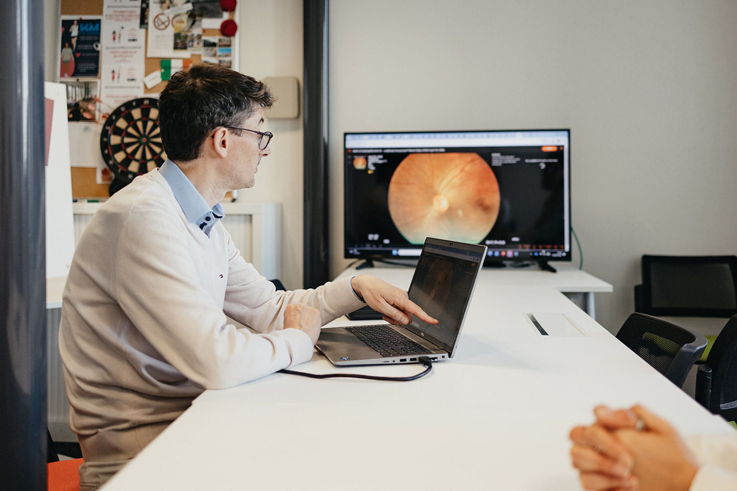
For more than 30 years, ADCIS has been developing computer vision, image analysis, and artificial intelligence solutions dedicated to ophthalmology. Our technologies automatically transform ophthalmic images into quantitative clinical data, helping eye care professionals, researchers, and industry partners improve efficiency, standardization, and diagnostic confidence. From retinal screening and corneal assessment to ocular surface analysis and lens design, ADCIS delivers innovative software solutions that support the entire ophthalmic care pathway.
By combining scientific expertise, clinical collaboration, and AI innovation, ADCIS helps transform ophthalmic images into actionable insights that improve patient care and accelerate research.
Our Expertise in Ophthalmology
ADCIS develops advanced image analysis and AI solutions across the entire eye :

Corneal image Analysis
AI-powered platform for in-vivo corneal tissue assessment based on speculare imaging

Retinal Imaging
AI-based screening of major retinal pathologies from color fundus images

Eye Surface
Eye redness automatic grading

Contact Lens
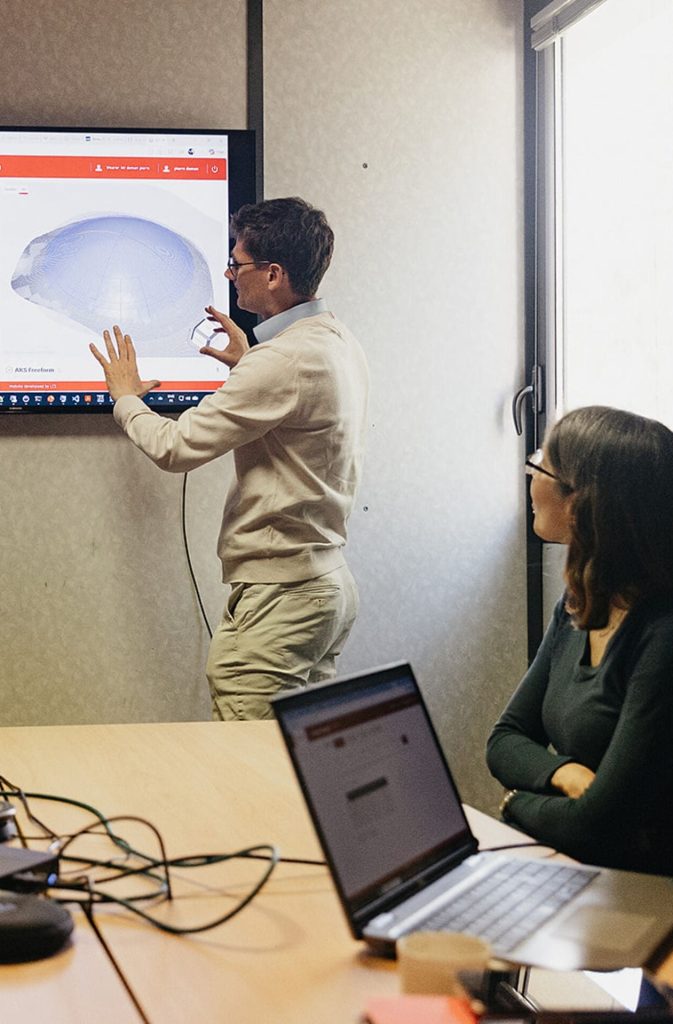
Computer-assisted fitting of custom scleral lenses based on patient-specific corneal profile
Your partner in
Image Analysis
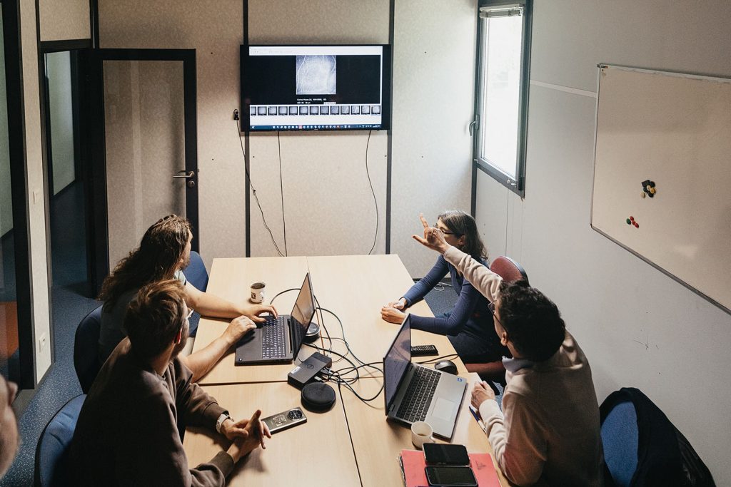
ADCIS collaborates with ophthalmologists, research laboratories, universities, hospitals, pharmaceutical companies, CROs, and medical device manufacturers to develop innovative ophthalmic imaging solutions. Its software products are co-developed with clinical and academic partners and are used in routine care, research projects, and clinical studies.
Our expertise supports :
- Eye care professionals
- Research laboratories and academic institutions
- Pharmaceutical and Medtech companies
- Clinical research organizations (CROs)
- Medical device manufacturers
- Eye banks and transplant centers
Whether for clinical routine, clinical trials, medical device development, or ophthalmic research, ADCIS provides AI-powered solutions that transform ophthalmic imaging into measurable clinical value.

Real-World Applications
- Diabetic Retinopathy Screening
- AMD & Glaucoma Monitoring
- Endothelial Cell Analysis
- Fuchs Dystrophy Assessment
- Corneal Graft Qualification
- Eye Bank Quality Control
- Dry Eye Evaluation
- Ocular Redness Quantification
- Custom Scleral Lens Design
- Clinical Trial Imaging Endpoints
AI-Powered Imaging for Better Eye Care
Healthcare professionals need reliable and reproducible tools to analyze increasingly complex ophthalmic images. Manual interpretation can be time-consuming, subjective, and difficult to standardize across clinical centers and studies.
ADCIS solutions help organizations :
- Improve diagnostic accuracy
- Standardize image interpretation
- Automate quantitative measurements
- Support clinical decision-making
- Accelerate ophthalmic research
- Generate reproducible imaging endpoints
- Facilitate patient monitoring over time
- Improve efficiency in clinical workflows
Our software solutions are trusted by clinicians, researchers, and device manufacturers worldwide to deliver objective and reliable ophthalmic image analysis.
Messidor, the retinal image database
dedicated to research
ADCIS hosts the image database from the Messidor project (Méthodes d’Évaluation de Systèmes de Segmentation et d’Indexation Dédiées à l’Ophtalmologie Rétinienne). Developed to support studies on computer-assisted diagnosis of diabetic retinopathy, the Messidor database consists of 1,200 color fundus images and is available to the public.
Let’s Discuss Your Projects !
Discover how ADCIS computer vision and AI solutions can improve diagnosis,
accelerate research, and deliver objective ophthalmic image analysis.
Book a call with an ADCIS expert – and let’s find out how we can accelerate your next imaging project.
Email: adcis-team@adcis.net
Phone: (+33)2 31 06 23 00
Address: 3 rue Martin Luther King 14280 SAINT-CONTEST – FRANCE